Breast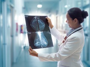 cancer is one of the most prevalent and concerning health issues faced by women worldwide. Early detection plays a crucial role in improving survival rates and treatment outcomes. Fortunately, medical imaging techniques have revolutionized breast cancer screening, diagnosis, and monitoring. In this blog post, we will delve into the world of medical imaging for women’s health, specifically focusing on breast cancer screening and exploring the broader applications of medical imaging beyond breast cancer detection. Breast cancer is a malignant condition that occurs when abnormal cells in the breast tissue multiply uncontrollably. It can develop in women of any age, although the risk increases with age. Regular screening is vital for early detection because early-stage breast cancer is often asymptomatic. Medical imaging offers valuable tools for breast cancer screening. Mammography is the most common imaging technique used, especially for women aged 40 and older. It employs low-dose X-rays to create detailed images of the breast tissue, allowing radiologists to identify any suspicious masses or calcifications. Digital mammography, 3D mammography (tomosynthesis), and contrast-enhanced mammography are some advanced variations that provide enhanced visualization and accuracy. While mammography remains the gold standard, there are other imaging modalities that complement or supplement breast cancer screening. These include ultrasound and magnetic resonance imaging (MRI). Ultrasound tests utilize sound waves to generate real-time images of the breast tissue. It is particularly useful for distinguishing between solid masses and fluid-filled cysts. Ultrasound is also commonly employed to guide biopsies, aiding in the collection of tissue samples for further analysis. MRI, on the other hand, employs a powerful magnetic field and radio waves to create highly detailed images of the breast. It is recommended for women with a high risk of breast cancer or those with dense breast tissue. MRI provides enhanced sensitivity in detecting tumors, but it is often used as a supplementary tool due to its higher cost and longer imaging time.
Medical Imaging for Breast Cancer Diagnosis and Staging
In addition to screening, medical imaging plays a crucial role in diagnosing and staging breast cancer. If an abnormality is detected during screening or if a woman experiences symptoms such as a lump or nipple discharge, further imaging tests may be conducted. Diagnostic mammography provides more detailed images of specific areas of concern identified during screening. It allows radiologists to assess the nature of the abnormality and determine the need for additional tests or a biopsy. Biopsy, often guided by medical imaging, is a critical procedure for confirming breast cancer. Imaging techniques like ultrasound, MRI, or stereotactic mammography help locate the precise area for tissue sampling, ensuring accuracy and reducing invasiveness. Medical imaging also aids in breast cancer staging, which determines the extent and spread of the disease. Imaging techniques such as MRI, positron emission tomography (PET), and computed tomography (CT) scans are used to evaluate the presence of metastases in lymph nodes and other parts of the body.
Medical imaging has transformed breast cancer screening, diagnosis, and staging, contributing significantly to improved outcomes for women’s health. Mammography, ultrasound, and MRI serve as powerful tools in early detection, differentiation of benign and malignant lesions, and guiding biopsies. These imaging modalities play a vital role not only in breast cancer but also in monitoring treatment response and detecting potential recurrences. As technology advances, medical imaging continues to evolve, offering enhanced accuracy, improved patient comfort, and more efficient diagnoses. Regular screenings and appropriate imaging techniques empower women in the fight against breast cancer, enabling early interventions and ultimately saving lives. While breast cancer screening and diagnosis are prominent applications of medical imaging in women’s health, these imaging techniques have broader implications. For example, medical imaging plays a crucial role in evaluating and diagnosing other breast conditions, such as fibroadenomas, cysts, and mastitis. It helps differentiate between benign and malignant lesions, aiding in the development of appropriate treatment plans. Fibroadenomas, which are common benign breast tumors, can be accurately identified using ultrasound or mammography. These imaging techniques can provide valuable information about the size, shape, and characteristics of fibroadenoma, helping physicians determine the need for further intervention or monitoring.
Cysts, fluid-filled sacs that can develop in the breast tissue, are another condition where medical imaging plays a vital role. Ultrasound imaging is particularly useful in identifying and characterizing breast cysts. The imaging allows physicians to determine whether a cyst is simple or complex, guiding decisions about follow-up or intervention. Mastitis, an infection of the breast tissue that often occurs during breastfeeding, can also be evaluated using medical imaging. Ultrasound imaging can help identify the presence of abscesses, guide appropriate drainage procedures, and monitor the effectiveness of treatment.
Medical imaging is also essential for monitoring treatment response and detecting potential recurrences. After breast cancer diagnosis and initiation of treatment, regular imaging scans are conducted to assess the effectiveness of therapies, such as chemotherapy or radiation. Follow-up mammography, ultrasound, or MRI scans help evaluate tumor size, detect changes in breast tissue, and provide early indications of disease progression or relapse. Early detection of recurrent breast cancer enables timely intervention and can significantly impact treatment outcomes.
Moreover, medical imaging techniques find application in various gynecological conditions. Ultrasound is commonly used for assessing pelvic abnormalities, monitoring ovarian cysts, and evaluating the reproductive organs. It is a non-invasive imaging modality that provides detailed visualization of the pelvic structures, including the uterus, ovaries, and fallopian tubes. Ultrasound can help identify conditions such as polycystic ovary syndrome (PCOS), uterine fibroids, and ovarian tumors. It also plays a crucial role in guiding procedures like hysterosonography where saline is introduced into the uterus to enhance imaging and evaluate abnormalities in the uterine cavity.
MRI is another powerful tool employed in women’s health. It provides detailed images of the pelvis, offering valuable information about the uterus, ovaries, and surrounding structures. MRI is particularly useful in detecting and characterizing uterine fibroids, noncancerous growths that can cause pain, heavy menstrual bleeding, and other symptoms. It helps assess the size, location, and vascularity of fibroids, aiding in treatment planning and decision-making. In the field of infertility, medical imaging techniques contribute to the evaluation of causes and treatment planning. Transvaginal ultrasound allows visualization of the ovaries, follicles, and endometrial lining, providing crucial information about the menstrual cycle and ovulation. It helps diagnose conditions like polycystic ovary syndrome (PCOS) and assess the response to fertility medications. Furthermore, medical imaging plays a role in the diagnosis and evaluation of endometriosis, a condition where the tissue lining the uterus grows outside of it. MRI imaging can help identify and map the extent of endometrial implants, guiding treatment decisions and surgical planning. Medical imaging is also valuable in assessing pelvic pain causes in women. Ultrasound and MRI can help identify conditions such as ovarian cysts, pelvic inflammatory disease (PID), adhesions, and tumors. These imaging techniques provide visual information to aid in the diagnosis and management of pelvic pain, enabling targeted treatment approaches.
Medical imaging has revolutionized the field of women’s health by providing valuable tools for breast cancer screening, diagnosis, and monitoring. Beyond breast cancer, medical imaging techniques find broad applications in evaluating and diagnosing various breast conditions, such as fibroadenomas, cysts, and mastitis. They contribute to treatment monitoring and the early detection of recurrences. In gynecology, medical imaging plays a crucial role in assessing pelvic abnormalities, evaluating ovarian cysts, and diagnosing conditions like PCOS, uterine fibroids, and endometriosis. It aids in infertility evaluations, providing information about the menstrual cycle, ovulation, and response to fertility treatments. Furthermore, medical imaging assists in the diagnosis and management of pelvic pain causes, guiding targeted treatment approaches. As technology advances, medical imaging continues to evolve, offering enhanced accuracy, improved patient comfort, and more efficient diagnoses. The expanded applications of medical imaging in women’s health empower both patients and healthcare providers, enabling comprehensive evaluations, personalized treatment plans, and better outcomes. With ongoing advancements and research, medical imaging will continue to play a pivotal role in women’s health, contributing to early detection, effective treatment, and improved quality of life.
Breast cancer screening and diagnosis are vital aspects of women’s healthcare, but medical imaging has a broader range of applications beyond breast cancer. In this section, we will explore additional areas where medical imaging plays a crucial role in women’s health, providing valuable diagnostic information and guiding treatment decisions.
Obstetrics and Prenatal Care:
Medical imaging techniques are instrumental in monitoring the health and development of the fetus during pregnancy. Ultrasound imaging is the primary modality used in obstetrics. It allows healthcare providers to assess fetal growth, identify any abnormalities, and evaluate the well-being of both the mother and the baby. Ultrasound can determine gestational age, detect multiple pregnancies, and screen for conditions like fetal anomalies, placental abnormalities, and ectopic pregnancies. It also plays a crucial role in guiding procedures such as amniocentesis or chorionic villus sampling (CVS) for genetic testing.
In high-risk pregnancies, additional imaging techniques may be employed. Doppler ultrasound measures blood flow in the umbilical cord and placenta, aiding in the assessment of fetal well-being. Magnetic resonance imaging (MRI) may be used in specific cases where further evaluation is necessary, providing detailed anatomical information about the fetus and surrounding structures.
Pelvic Floor Disorders:
Pelvic floor disorders, including pelvic organ prolapse, urinary incontinence, and fecal incontinence, are common conditions that affect many women. Medical imaging techniques, such as pelvic ultrasound and MRI, are valuable tools in diagnosing and assessing these disorders. Ultrasound can evaluate the position and integrity of pelvic organs, such as the bladder and uterus, and assess the degree of prolapse. It can also provide real-time information about the function of the pelvic floor muscles during activities such as coughing or bearing down. MRI is particularly useful in providing detailed anatomical information and assessing the extent of pelvic organ prolapse. It can help identify contributing factors, such as changes in the position and support structures of the pelvic organs, allowing for personalized treatment planning.
Osteoporosis and Bone Health:
Osteoporosis is a condition characterized by decreased bone density and an increased risk of fractures. Medical imaging, specifically dual-energy X-ray absorptiometry (DXA), is the gold standard for assessing bone mineral density and diagnosing osteoporosis. DXA scans provide precise measurements of bone density in key areas such as the hip and spine, enabling early detection and monitoring of the disease. These measurements are essential in assessing fracture risk and guiding treatment decisions.
Cardiovascular Health:
Cardiovascular disease is a leading cause of mortality in women. Medical imaging plays a vital role in the evaluation and management of cardiovascular conditions. Techniques such as echocardiography, cardiac CT scans, and cardiac MRI provide detailed information about the structure and function of the heart, helping diagnose conditions such as heart disease, valve disorders, and heart failure. These imaging modalities assess cardiac function, visualize blood flow, and identify any abnormalities or blockages in the arteries, contributing to effective treatment planning.
Why Choose Hollywood Diagnostics for Women’s Health Imaging in South Florida?
When it comes to women’s health imaging, choosing the right medical imaging center is of utmost importance. For women in South Florida, Hollywood Diagnostics stands out as a premier choice. Here are some compelling reasons why women should consider Hollywood Diagnostics for their imaging needs. Hollywood Diagnostics is staffed with a team of highly skilled and experienced radiologists who specialize in women’s health imaging. These experts have extensive knowledge and training in interpreting breast imaging, obstetric ultrasound, gynecological imaging, and other related modalities. Their expertise ensures accurate diagnoses and personalized care for every patient.
State-of-the-Art Technology:
- Hollywood Diagnostics is equipped with the latest and most advanced medical imaging technology. The center invests in state-of-the-art equipment, such as digital mammography, 3D tomosynthesis, high-resolution ultrasound machines, and MRI scanners with specialized coils for women’s health imaging. This cutting-edge technology provides detailed and high-quality images, enhancing diagnostic accuracy and improving patient outcomes.
Comprehensive Range of Services:
- Hollywood Diagnostics offers a comprehensive range of imaging services tailored specifically to women’s health needs. From breast cancer screening and diagnosis to prenatal ultrasound, pelvic floor imaging, and evaluation of gynecological conditions, the center provides a full spectrum of imaging services. This comprehensive approach ensures that women receive all the necessary imaging evaluations under one roof, streamlining the diagnostic process and facilitating continuity of care.
Patient-Centered Approach:
- At Hollywood Diagnostics, patient comfort, safety, and well-being are top priorities. The center is designed to create a warm and welcoming environment, with dedicated waiting areas and private examination rooms. The staff takes extra care to ensure patient comfort during procedures and strives to address any concerns or questions that patients may have. The radiologists and technologists work closely with patients, explaining the imaging process, addressing anxieties, and providing support throughout the examination.
Timely and Efficient Service:
- Hollywood Diagnostics understands the importance of timely results in women’s health imaging. The center is committed to providing fast turnaround times for imaging studies, ensuring that patients receive their results promptly. This efficiency is crucial in cases where further intervention or treatment planning may be required. The center also offers convenient scheduling options, including same-day appointments and extended hours, to accommodate busy schedules and urgent cases.
Collaborative Approach to Care:
- Hollywood Diagnostics emphasizes collaboration and communication with referring physicians and healthcare providers. The radiologists work closely with the patient’s healthcare team, providing detailed reports and actively participating in multidisciplinary discussions. This collaborative approach ensures seamless coordination of care, facilitates informed decision-making, and enhances overall patient management.
Women in South Florida can trust Hollywood Diagnostics for their women’s health imaging needs. With a team of expert radiologists, state-of-the-art technology, comprehensive services, patient-centered care, efficient service, and a collaborative approach, Hollywood Diagnostics is dedicated to delivering the highest quality of care and improving the health outcomes of women in the community.